Prof. Dr. Sabine Eming

Head of Research Area 2, Department of Dermatology, Faculty of Medicine

Prof. Sabine Eming CECAD

Prof. Sabine Eming

Head of Research Area 2, Principal Investigator

+49 221 478-3196

sabine.eming[at]uni-koeln.de

Klinik und Poliklinik für Dermatologie und Venerologie

Uniklinik Köln
Kerpener Str. 62

50937 Köln

Profile

Age-related decline in skin function is a main cause of morbidity and a major risk factor for infections, skin fragility, impaired wound healing, and skin cancer. Sabine Eming’s team is investigating the molecular basis of age-related skin pathologies. A major goal is to translate these findings into new diagnostic tools and therapeutic concepts.

Our research:
The wound healing response is essential to restore the skin barrier and its functions rapidly after injury. The efficiency and the quality of the repair response vary considerably during a person’s lifetime. Whereas fetal skin is characterized by perfect skin regeneration, postnatal skin damage is replaced by a scar, and wound healing is delayed with increasing age. In fact, aging is a significant risk factor for chronic wounds. Incomplete understanding of the underlying molecular basis of tissue repair and its failure is the main reason for the lack of efficient means of treating non-healing wounds and reducing scar formation. The field of tissue repair and regeneration therefore has great potential for tackling one of the great health concerns and needs of the aging population. The Eming lab aims to identify the molecular networks controlling wound healing and its failure. But wound healing mechanisms are not unique to the tissue repair response. Postnatal wound healing in part recapitulates processes in developmental biology and organogenesis. Signals controlling cell growth, migration, and differentiation during tissue repair have also emerged as central mediators in cancer biology and inflammatory disease processes. Prof. Eming and her team are focusing on the cellular and molecular principles of tissue repair as a model for studying interrelations between the aging-related decline of regenerative capacity and aging-associated pathological skin conditions, including cancer and inflammation. The group is particularly interested in identifying functional links between mechanisms that suppress wound healing ability and promote skin cancer as the body ages.

Our successes:
There is substantial evidence for an inverse correlation between immune competence and the tissue’s capacity to regenerate or repair itself by forming a scar. Studies in mammals with limited regenerative capacity and findings in non-mammalian model organisms with a high regenerative capacity have contributed to this paradigm. In the past decade, the Eming lab has made seminal contributions to unraveling the mechanisms by which the immune system intervenes in the healing response or its failure.
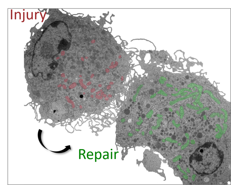
 Using a systems biology approach in wound tissue of mice and humans, they have identified novel inflammatory proteases and targets in the wound environment and provided evidence on how their interactions disturb downstream cell functions essential for repair. These studies have led to the discovery of new molecular control mechanisms for proteins of the VEGF family and furthered understanding of the role of VEGF proteins and their receptors in vessel formation and tissue regeneration. The findings have provided insight into how engineered growth factors in concert with biomaterials may offer a potent molecular approach for therapeutic angiogenesis. The group has also developed new genetically modified mouse models and techniques to investigate how monoytes/macrophages repair skin wounds and how these cells contribute to skin carcinogenesis. In collaboration with other investigators, they have recently identified new pathways that may underlie scarless repair in zebrafish but determine scar formation in mice and humans. The group has contributed to numerous clinical trials to optimize therapeutic strategies for conditions with impaired healing.

Our goals:
The Eming lab is aiming at deeper understanding of how cells sense tissue damage and how these events translate into a regenerative response or disease. The ultimate goal is to reprogram the healing response, in order to readjust postnatal repair into regeneration and to develop novel strategies for pharmacological interventions to facilitate healing of injured, diseased or aging tissues. The close interaction between basic science and clinical expertise enables the group to translate new findings into therapeutic approaches.

Our methods/techniques:
Prof. Eming leads a program of research into tissue damage and repair encompassing basic structure/function analysis, in vivo models, and human disease. The group has longstanding expertise in cell-cell and cell-matrix interactions relevant to various aspects of tissue remodeling and repair. A wide range of in vitro and in vivo models allows the team to analyze the function and crosstalk of diverse types of skin cell. Methods include a number of cell systems (monolayer cultures, complex co-cultures), as well as genetically modified mouse models and preclinical disease models combined with proteomics, gene expression analysis, and imaging techniques.

Figures

Figure 1: Identification of plasmin cleavage sites within the Heparin binding domain of VEGF-A165 and PLGF-1/-2, and generation of novel proteolysis resistant VEGF mutants with pro-angiogenic activity. A) VEGF-A165 and PLGF-1/-2 degradation products were isolated in exsudates obtained from human non-healing Venous leg ulcers. Plasmin cleavage sites were idientified by LC-MS/MS analysis. B) Site directed mutagenesis substituted Arg110 with Ala110 in VEGF-A165, yielding a plasmin-resistant VEGF-A mutant with higher stability and binding affinity to heparin sulfate proteoglycans in the chronic wound environment which ultimately improved the pro-angiogenic activity.

Figure 2: Engineering the growth factor microenvironment with fibronectin domains to promote wound healing. A) Generation of a novel bi-functional protein (FNIII10-VEGF) consisting of the 10th type III domain of fibronectin (FNIII10) fused to VEGF-A165 (VEGF) with potentiated angiogenic activity. FNIII10-VEGF activates simultaneously VEGF-receptor 2 and αvβ3 integrins in endothelial cells resulting in synergistic downstream effects. B) FNIII10-VEGF promotes cell spreading, clustering of αvβ3 integrins and Rac1 activation.

Figure 3: Macrophages are the prevailing VEGF-A source during the early wound healing phase. A) X-Gal staining of unwounded and wounded skin of VEGF-lacZ reporter mice. B) X-Gal+ wound cells were isolated and analyzed by FACS. A fraction of approximately 20% of all macrophages at the wound site expresses VEGF-A.

EXTERNAL Cooperations
  • Prof. Dr. Leptin, Genetic Institute, University of Cologn, DE