In no other organ can we see the aging process as well as in our skin. With age, the regenerative capacity declines, leading to impaired barrier function and wound healing. Dr. Parisa Kakanj conducts research in the group of Professor Maria Leptin and wants to understand the underlying mechanisms in skin cells. In particular, the focus is on the signaling pathways of food perception, insulin and TOR signaling, which have special roles in maintaining skin balance during aging and regeneration.
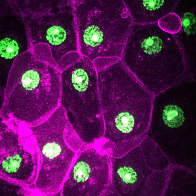
The image
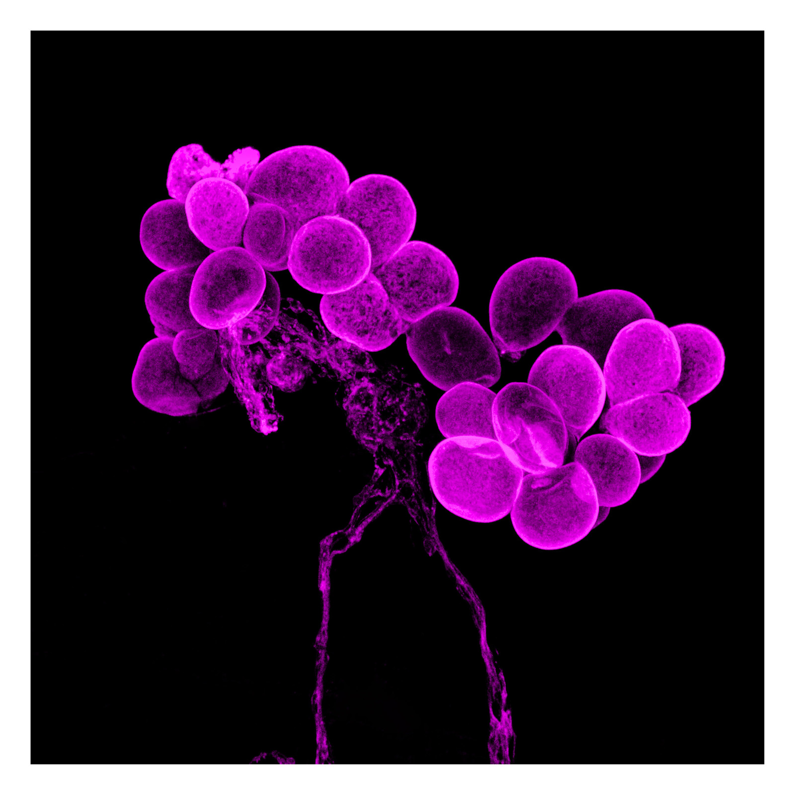
This image shows skin cells from a fly larva. The fruit fly Drosophila melanogaster is often used in science to understand basic mechanisms. This is possible because many genes are very similar between flies and humans. Here you can see faulty signaling in metabolism that causes large bubbles to form from cell membranes and drift away (magenta). Cell nuclei are in green.
The Photographer
Parisa Kakanj is a postdoctoral researcher in Professor Maria Leptin's lab. She has developed a method for live imaging and laser ablation of Drosophila larvae that is the basis for many interesting observations and results. "Having a challenging scientific question and observing phenomena for the first time is what drives me to be a scientist and fills me with great joy and enthusiasm," Kakanj said.
Fun Fact: If she is excited about an experiment, Parisa does a handstand in the lab.

Dr. Matthias Rübsam is fascinated by the barrier function of our skin, the epidermis, which, among other things, protects many animals from death by dehydration. Matthias is a scientist in the research group of Professor Carien Niessen.
Together, they are questioning how cells in the epidermis interact and self-organize to enable the formation and regeneration of this multi-layered barrier tissue. The epidermis comprises multiple cell layers with different tasks for each cell. Keratinocytes pass through all layers and tasks during their lifetime, from stem cell to fully transformed denucleated corneocyte, the corneal cell. How cell tasks and position within the tissue change over time and whether this is coupled is not clear.
The focus of the research group is how physical properties of cells, determined by intercellular junctions and the cytoskeleton, help position cells and define their task.

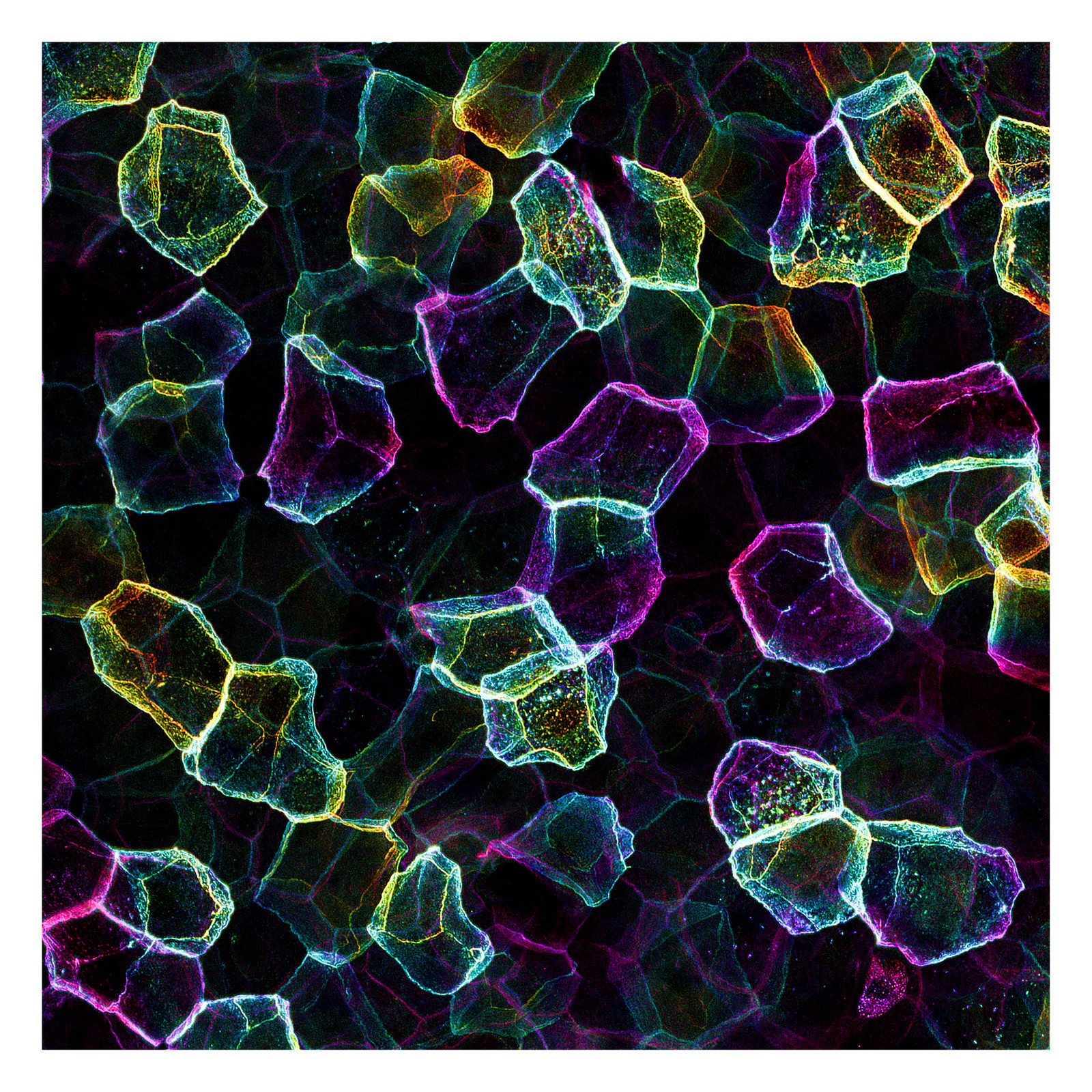
The image
Image shows pseudo-stained connections between skin cells and cytoskeleton (green) of keratinocytes in mouse skin.
The photographer
Matthias Rübsam is a scientist, a trained carpenter and petrolhead.
"I like to create and fix things, so I need to know how things made by humans or nature work. It turns out the problems caused by nature are harder to fix, so I stuck with those."
Fun Fact: Repairing cars is the most relaxing thing for me.
"Challenge Aging: Microcosm of Aging Research" is the title of our exhibition of microscopic images on the subject of aging. Aging challenges us all.
At the CECAD Excellence Cluster, we have accepted the challenge - and challenge aging itself with our research. We carry out basic research on various diseases, the risk of which increases with age: Cardiovascular diseases, cancer, Alzheimer's disease and diabetes. We are trying to answer the question of whether all these different diseases have a common basis.
Microscopy plays an important role in today's research. Thanks to state-of-the-art equipment and qualified staff, we are now able to gain insights into cells and organisms that were impossible just a few years ago.
As an outer protective layer, the skin prevents the unwanted intrusion of foreign substances into the human body and protects us from UV rays and overheating. Most importantly: without the protection that evaporation the skin offers, life on land would not be possible at all. The skin is made up of several layers in which different specialized cell types are located. These cell types interact with each other and exchange information via contact points in all spatial directions. Due to natural aging, the skin loses its elasticity over time, leading to wrinkle formation.
Picture: Dr. Christian Jüngst
Sample preparation: Dr. Matthias Rübsam (Niessen lab, CECAD)

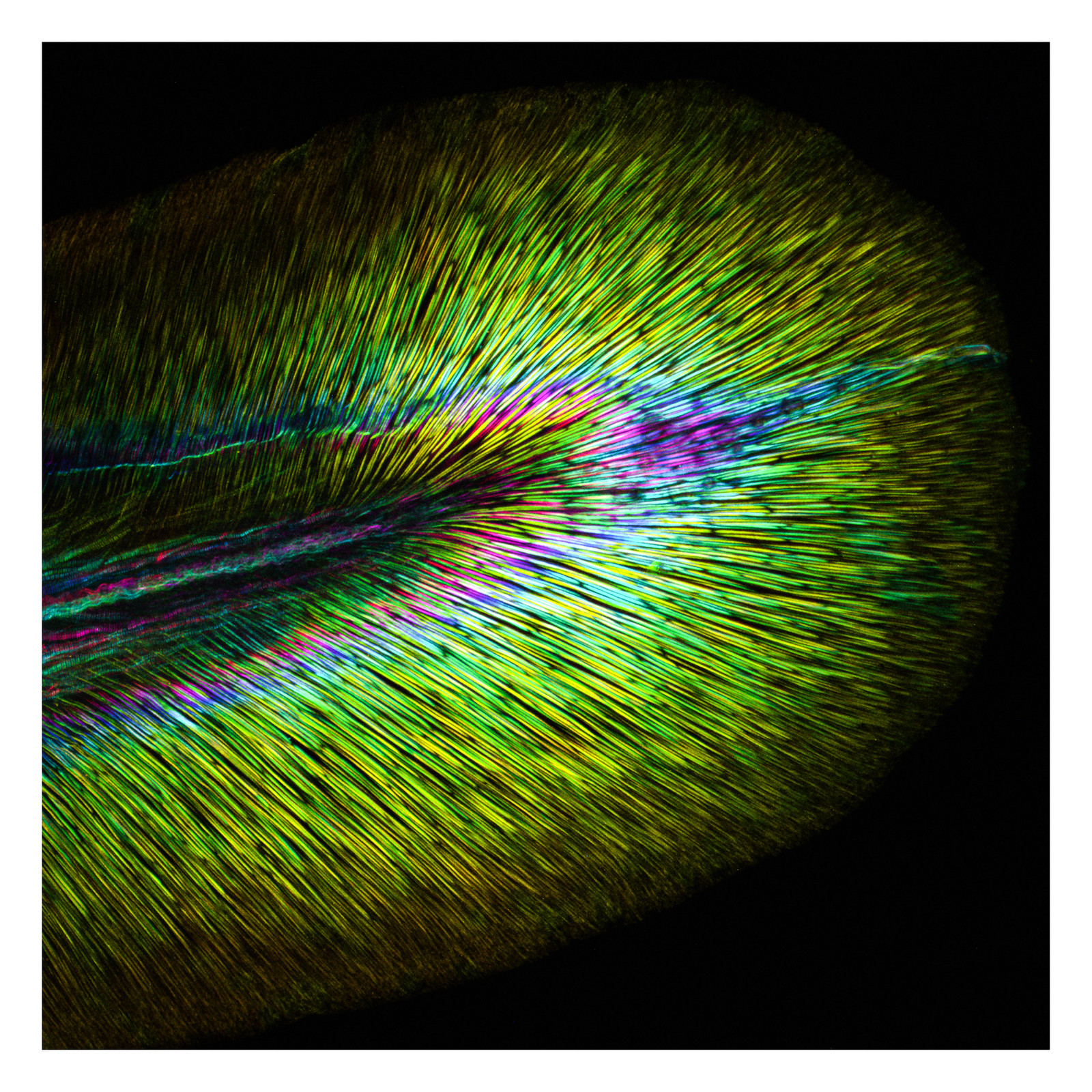
The musculature is an elementary component of most animals. It is indispensable for gross and fine motor activities and is often exposed to high loads. Here are shown symmetrical muscle fibres, as they extend over the entire trunk of a zebrafish embryo, to enable a directional locomotion. In order to prevent age-related muscle atrophy, the individual muscle areas should be trained continuously.
Picture: Dr. Christian Jüngst
Sample preparation: Dr. Daniela Weckler (Hammerschmidt lab, Zoologisches Institut der Universität zu Köln)
In order to ensure the optimal nutrient supply of all organs, they are traversed by many blood vessels, which can become clogged with increasing age. The widely branched blood vessels are stabilized by an enveloping layer of collagen fibres, which are visualized here with the aid of a label-free microscopy technique. In order to be able to look inside the tissue, the murine kidney used here was optically cleared by chemical treatment before the image was acquired.
Picture: Dr. Christian Jüngst
Sample preparation: Julia Binz (Nephrolab)
As the main driver-of locomotion, the tail fin is an indispensable part of the fish anatomy. With the help of its pronounced musculature (see also the picture Powerful Symmetry”) a fish can move its body wave-like along its axis, whereby a directed movement in the water becomes possible. This microscopic image of a zebrafish embryo shows the supporting elements of the tail fin, also known as actinotrichia, made of collagen fibres.
Picture: Dr. Christian Jüngst
Sample preparation: Dr. Daniela Weckler (Hammerschmidt lab, Zoologisches Institut der Universität zu Köln)
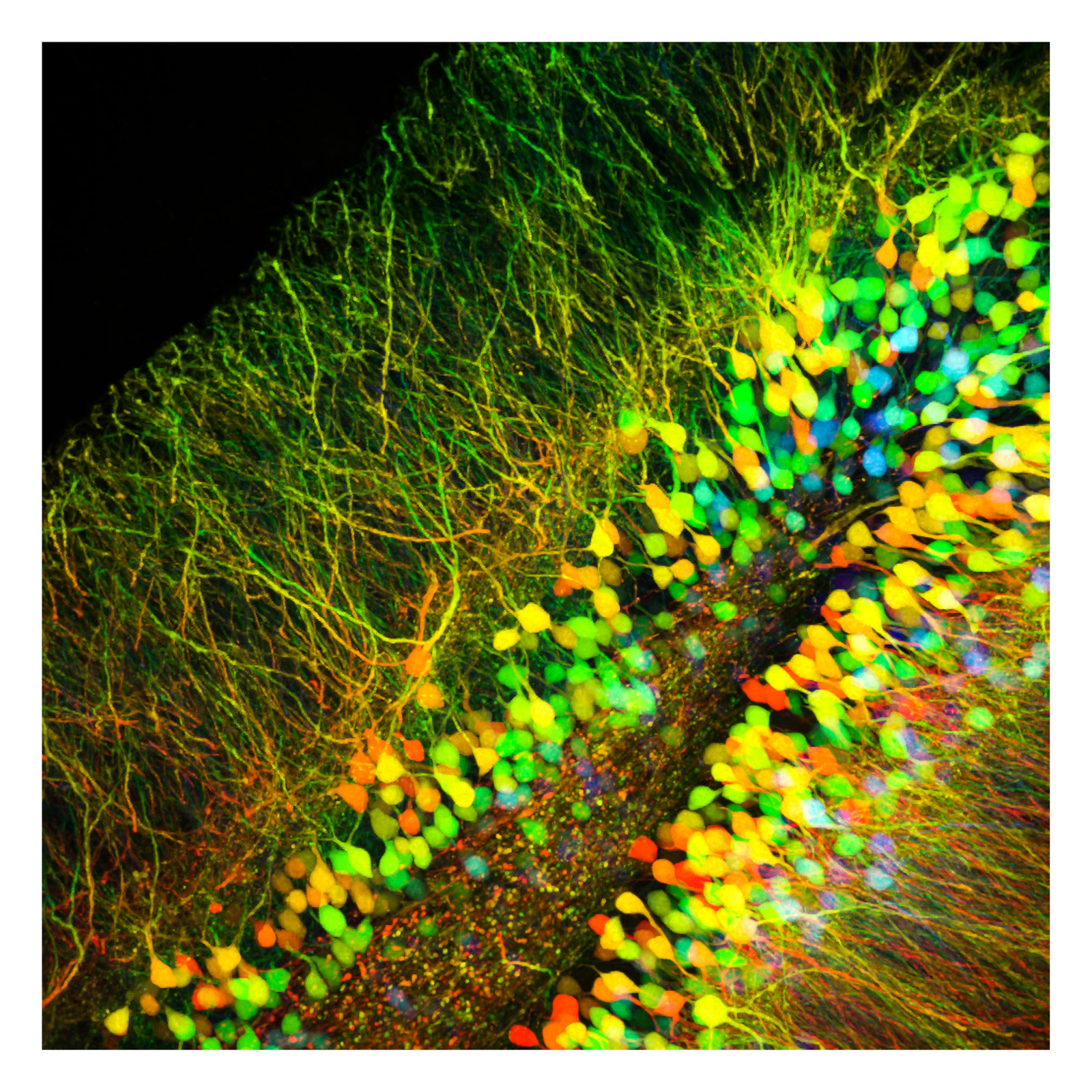
Many age-related diseases such as Alzheimer or Parkinson have their origin in the brain. The death of certain nerve cells leads to a disturbance of the complex neurophysiological processes and thus to neurodegenerative diseases. Genetically stained nerve cells, which are found billions of times in the brain, are shown here. Their interaction enables conscious control of the body and mental performance.
Picture: Dr. Christian Jüngst
Sample preparation: Bergami lab, CECAD
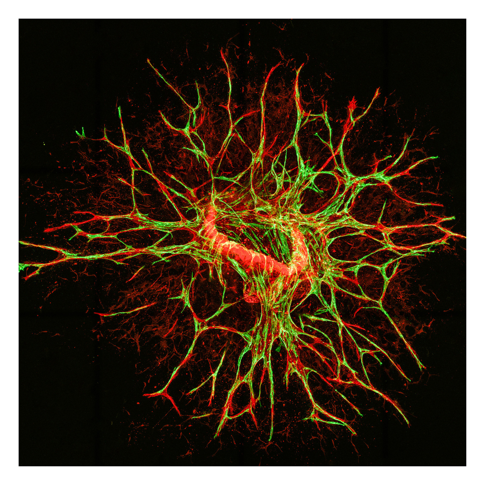
The formation or expansion of blood vessels is a decisive step in embryonic development, but also in regeneration processes such as wound healing. The efficiency can decrease with increasing age. In order to test the growth behaviour of the endothelial cells involved in vessel formation under different conditions, existing blood vessels can be embedded in a kind of gel in which the cells can spread in branched form in all directions.
Picture: Dr. Christian Jüngst
Sample preparation: Dr. Lars Schiffmann (Kashkar lab, CECAD)
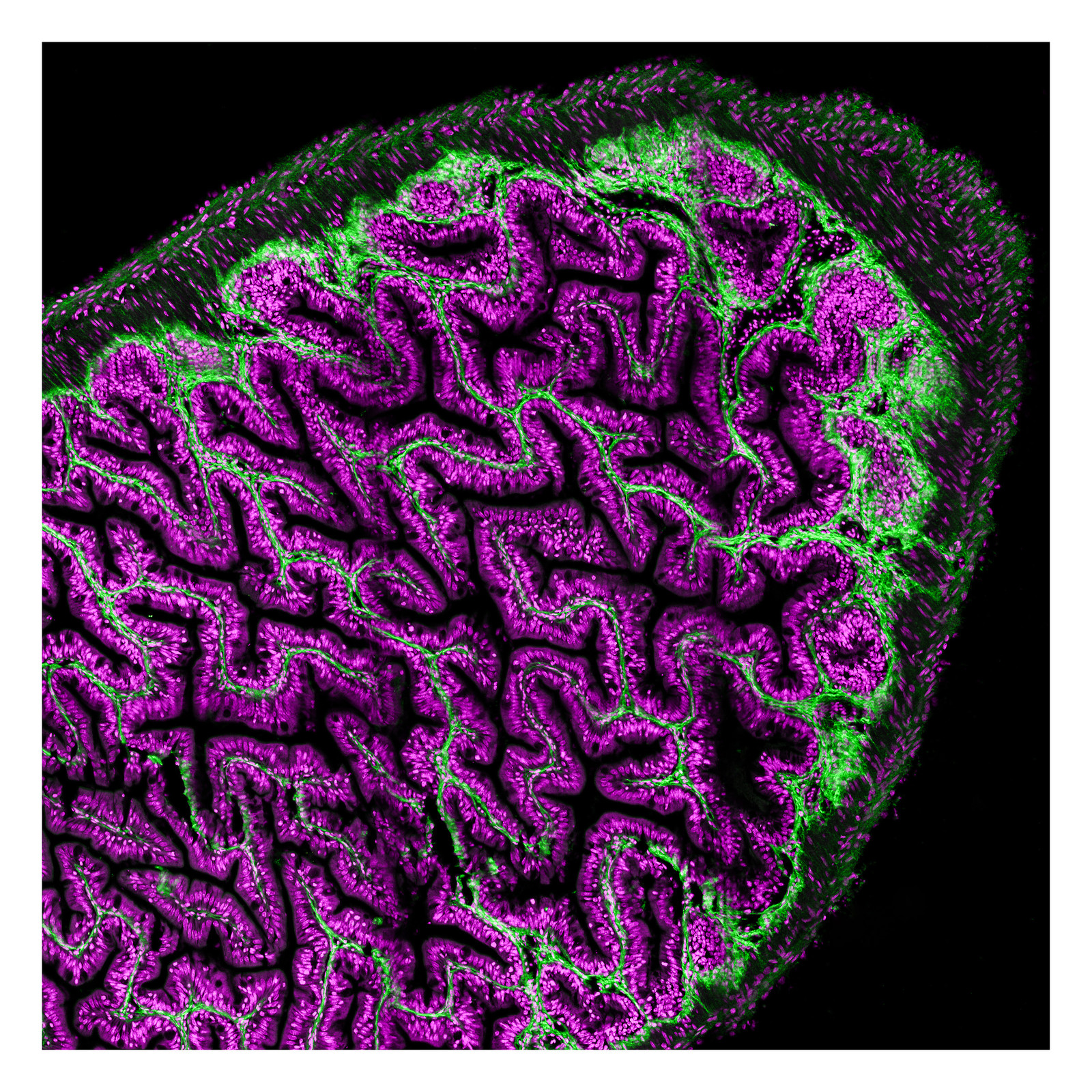
Killifishes play a special role in aging research. Unlike most other vertebrates, these small animals have a very short lifespan of only a few months, but show the same age-related diseases as long-living organisms. This offers clear advantages over the mouse as a classical model organism for aging research, as one does not have to wait several years to be able to carry out research on an old organism. The image shown here shows a cross-section of the fish intestine, which could be used, for example, to investigate age-related intestinal cancer.
Picture: Dr. Christian Jüngst
Sample preparation: Jens Seidel (Valenzano lab, MPI for Biology of Ageing)
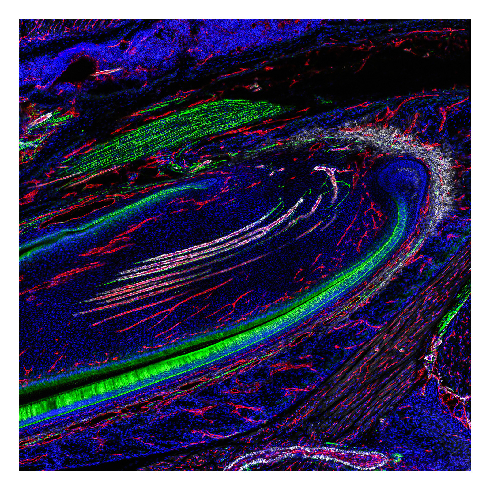
In rodents, the incisors renew and sharpen for the complete lifetime. Continuous tooth growth is made possible by stem cells and regulated by signals from nerves. This picture shows a longitudinal section of an incisor, with the nerves marked green, the blood vessels coloured red and smooth muscle cells in the vessel wall of arterioles appearing white.
Picture: Dr. Christian Jüngst
Sample preparation: Dr. Thomas Imhoff (Koch lab, Zentrum für Biochemie Köln)
The human kidney is indispensable for the excretion of metabolic waste products and toxins as well as for a balanced water and acid-base balance. As part of the natural aging process, the filtration performance of the kidneys, which is primarily ensured by specialized kidney cells (podocytes), decreases continuously. In order to understand the molecular mechanisms of the filtration process, model organisms such as the fruit fly Drosophila melanogaster are used. Drosophila has podocyte-like cells, the nephrocytes, which were stained here.
Picture: Johanna Odenthal (Brinkkötter lab, ZMMK)
Sample preparation: Johanna Odenthal (Brinkkötter lab, ZMMK)
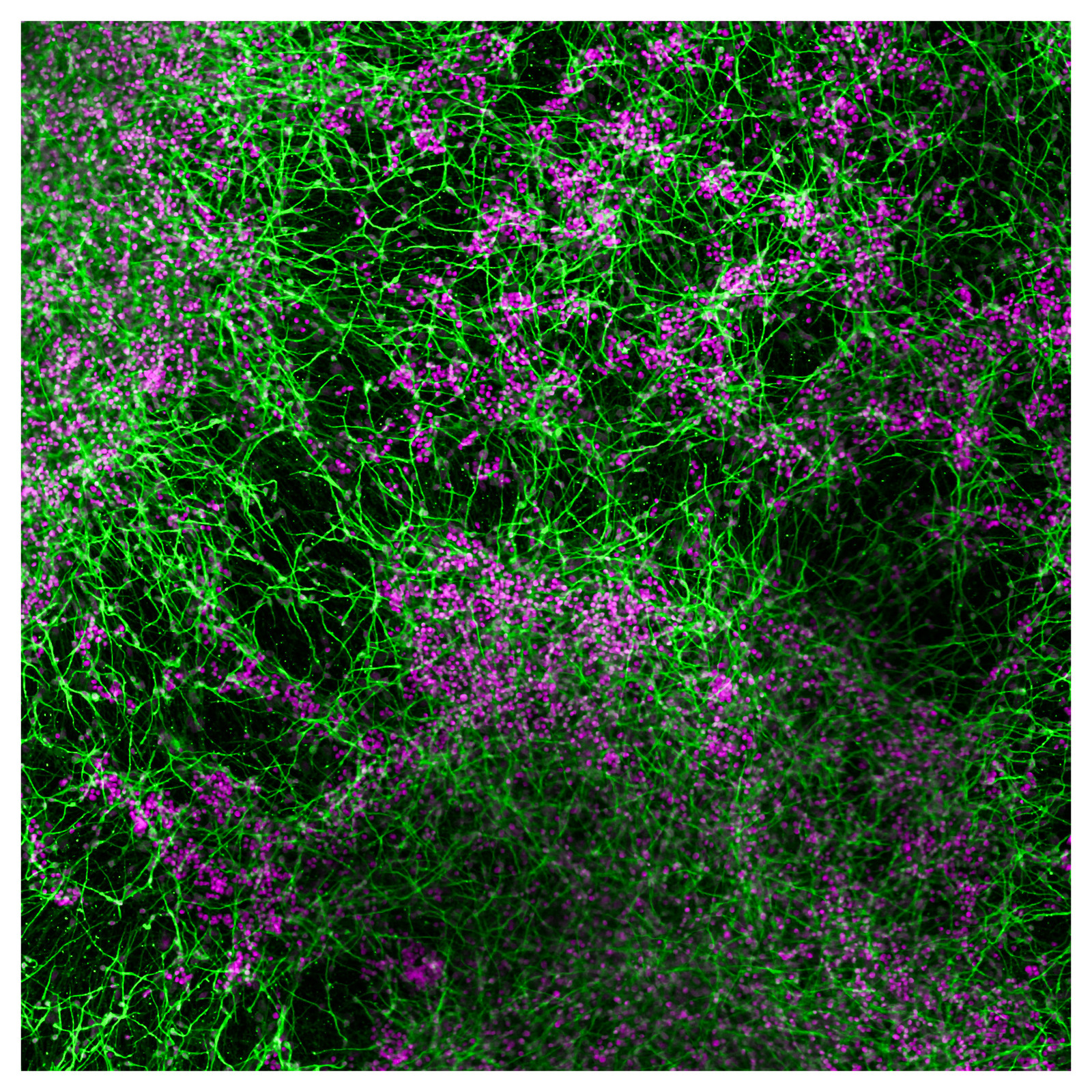
In a culture dish, stem cells can develop into neurons. Individual neurons, here shown in green, find and combine themselves to form their own cosmos, a network of thought. Each single purple point represents a cell nucleus. But not every cell nucleus can be assigned to a neuron, because there are other cells to discover in our brain cosmos.
Picture: Dr. Ines Lauria
Sample preparation: Dr. Ines Lauria and Farina Bendt (Fritsche lab, Leibniz Research Institute for Environmental Medicine, Düsseldorf)
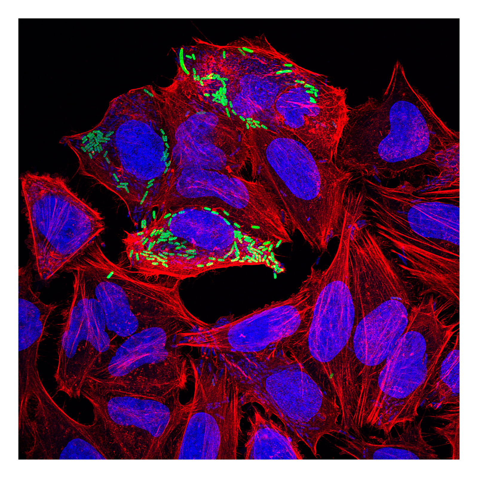
Infectious diseases are still one the major causes of death worldwide. One prominent representative is the enteroinvasive bacterium Shigella, which causes more than 1 million deaths every year. Shigella invades the intestinal epithelium and leads to severe bloody diarrhea. The image displays Shigella hijacking the host cytoskeleton to move throughout the cells. Due to this mechanism Shigella is able to evade the immune system, which patrols outside of the cells and would lead to the destruction of the bacterium.
Picture: Dr. Astrid Schauss
Sample preparation: Dr. Saskia Günther (Kashkar lab, CECAD)
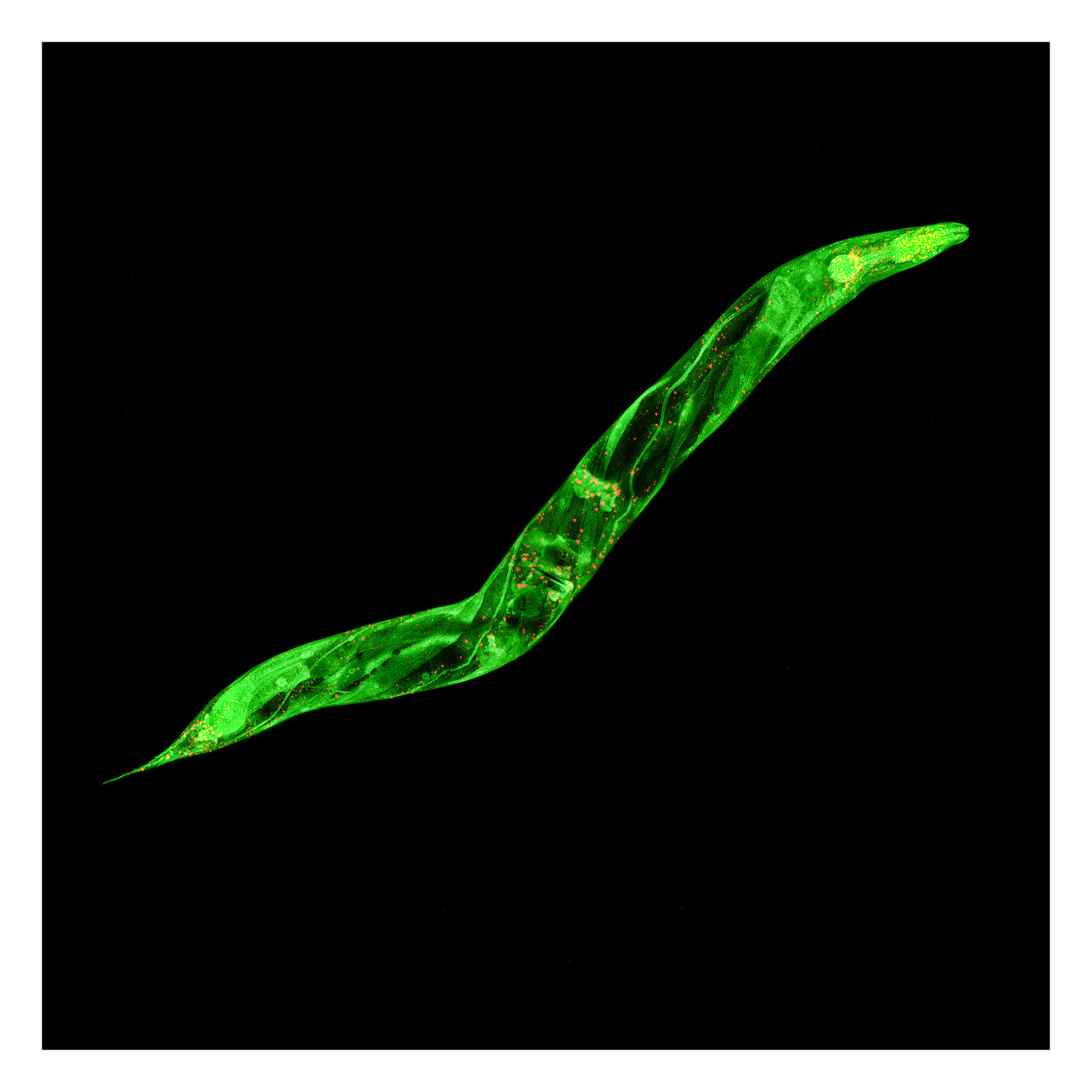
The picture shows a fluorescent image of a Caenorhabditis elegans, a nematode that is commonly used to study aging and to model age-related diseases. Two proteins are fluorescently labeled: The major protein synthesis factor eIF4E (shown in green) and the mRNA stability factor DCAP-1 (shown in red). Both factors play a central role in cellular protein production, stress response pathways and the aging process. Visualising these proteins in a living animal is of high relevance for understanding the molecular basis of illnesses, like Alzheimer’s disease or Parkinson’s disease.
Picture: Dr. Astrid Schauss
Sample preparation: Dr. Matthias Riecker (Schumacher lab, CECAD)
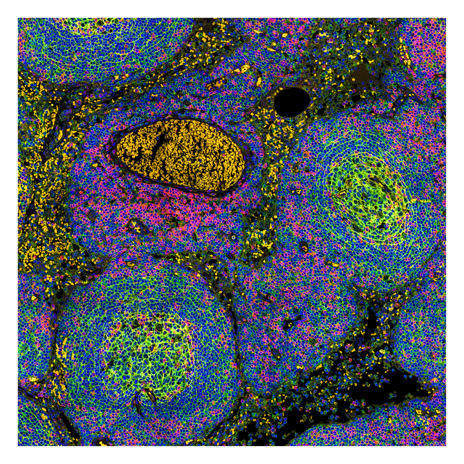
Lymph nodes do not only play a central role in the recognition of infections but also tumor diseases can be detected by the immune system. Lymphatic vessels span the whole body and are responsible for waste disposal of cell debris as well as intruded viruses, fungi or bacteria. Within the lymph nodes these particles are filtered out. The node also serves as a meeting point for different immune cells where they work tightly packed on a successful immune defence. The picture shows a section of a lymph node with an accumulation of differently stained immune cells.
Picture: Dr. Astrid Schauss
Sample preparation: M.Sc.Martin Thelen (Schlößer lab, ZMMK)
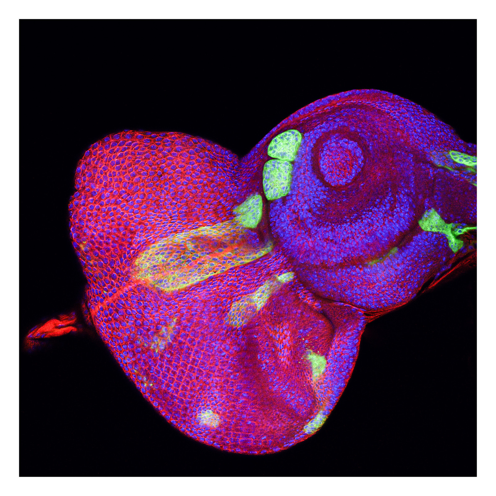
The development of our organs is a tightly regulated process. The fruit fly Drosophila melanogaster has been among the most favorite models to study development. Adult fly appendages such as eyes, legs or wings originate from special tissues, called imaginal discs. They have been used as a powerful model to study epithelial development, regeneration and cancer. The image depicts an eye/antennal imaginal disc, from which the composite eye and antenna will develop. The green color labels genetically reprogrammed cells, which form cyst-like structures.
Picture: Dr. Astrid Schauss
Sample preparation: Dr. Gábor Csordás (Uhlirova lab, CECAD)
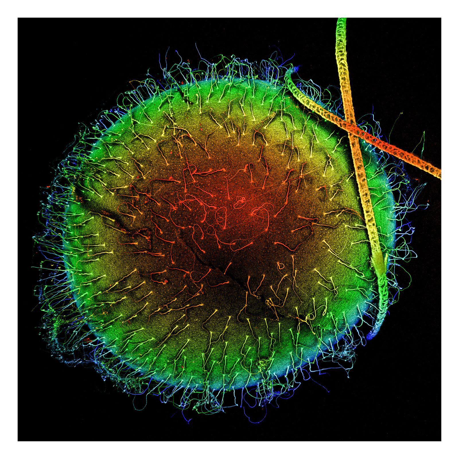
Turquoise killifish live in ephemeral freshwater pools on the African savannah, which form during the rainy season before quickly drying out. To survive in this harsh environment, they have evolved an fast life cycle in which fish are hatched and develop to maturity as quickly as two weeks, before laying eggs, which can lie dormant until the next rainy season. This makes them ideal models for aging research. The embryos are uniquely adapted to survive in this harsh desiccated environment, including a tough outer chorion, and hairlike filaments on the egg of so far unknown function.
Picture: Dr. Astrid Schauss
Sample preparation: Michael Poeschla (Valenzano lab, MPI for Biology of Ageing)
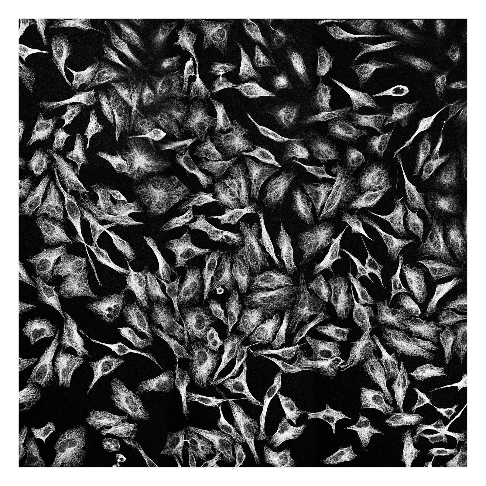
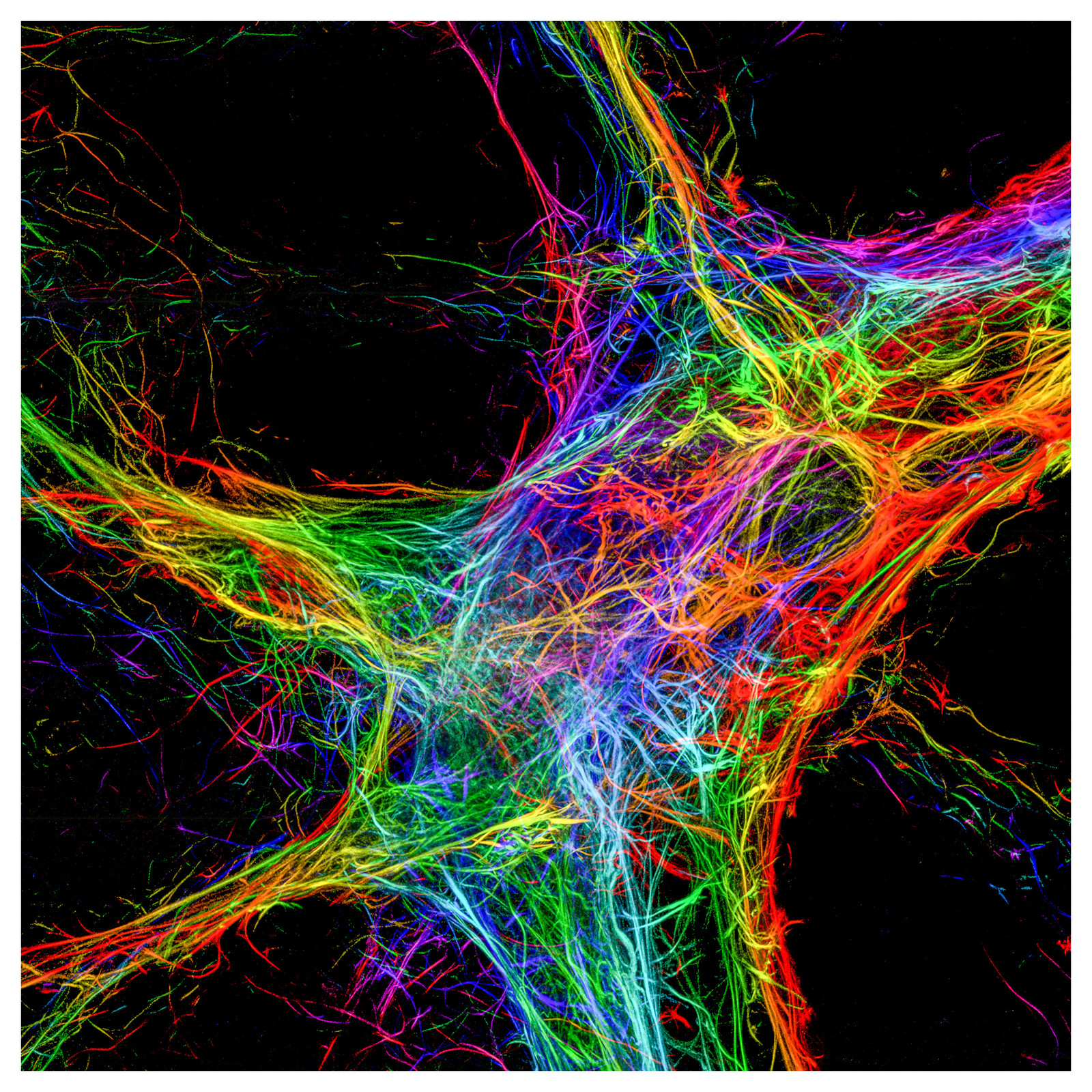
Cells in our body are using an inner cell skeleton to keep their shape. One of these components is tubulin which is fluorescently labelled in the image. Despite its role as structural protein, it is for example a key player in the segregation of chromosomes during cell division. As a cell´s railway system it is used to transport organelles and vesicles with different cargo, tracked by motor proteins as engines, to their destination.
Picture: Dr. Astrid Schauss
Sample preparation: Veronika Wulff (Schauss lab, CECAD)